Cytology questions | form five Biology
Find Cytology examination questions, form five Biology in acaproso.com
| # | Question | ||||||||||||
|---|---|---|---|---|---|---|---|---|---|---|---|---|---|
| 1 |
Long answers |
||||||||||||
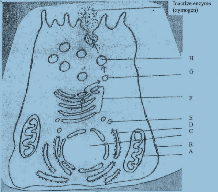
| 2 | Below is a diagram of an acinar cell of a pancreas involved in the synthesis and secretion of an inactive enzyme (zymogen)
Short answers |
||||||||||||
| 3 |
Short answers |
||||||||||||
| 4 |
Long answers |
||||||||||||
| 5 |
Short answers |
||||||||||||
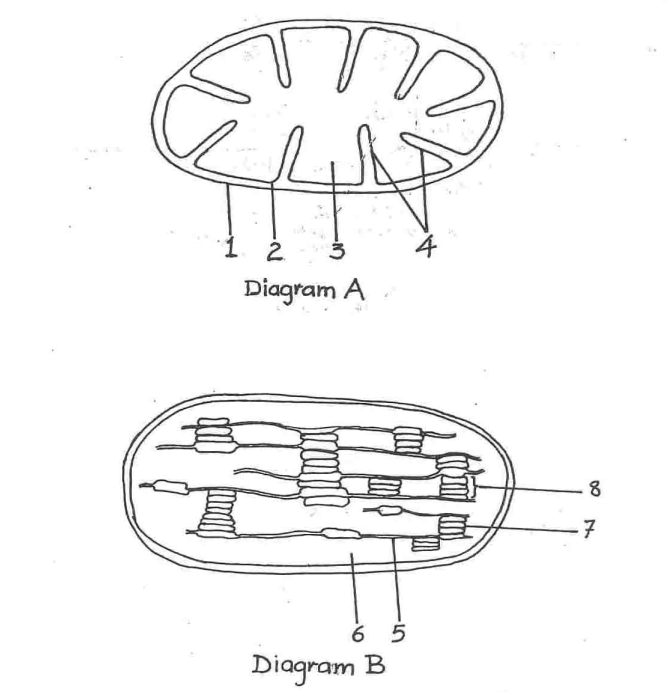
| 6 | Diagram A and B represent two cell organelles as seen under electronic microscope.
(ii) Name the structures numbered 1-8
Long answers |
||||||||||||
| 7 |
i) Golgi apparatus ii) Chloroplast
Long answers |
||||||||||||
| 8 |
Long answers |
||||||||||||
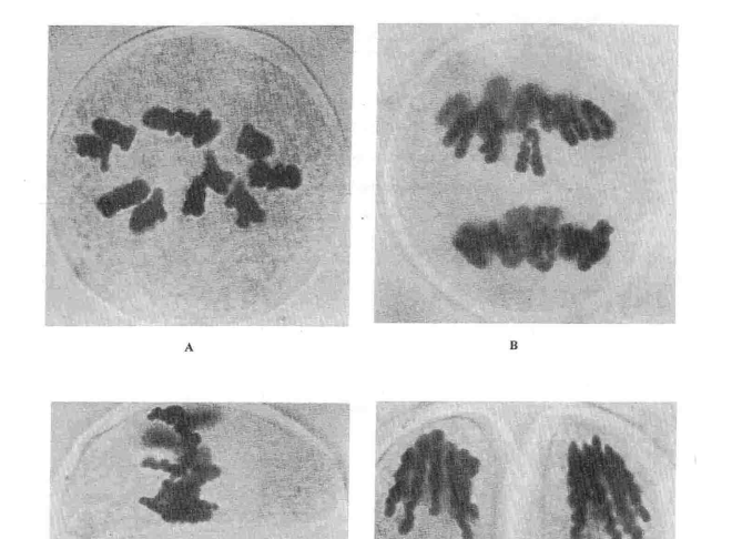
| 9 | The photomicrographs below; A, B, C and D swho different stages of animal cell undergoing division.
(ii) Describe the events taking place at each of the four stages in A, B, C and D
(ii)Where and when in a human being are likely to encounter this type of cell division?
Long answers |
||||||||||||
| 10 |
Short answers |
 Acaproso Login